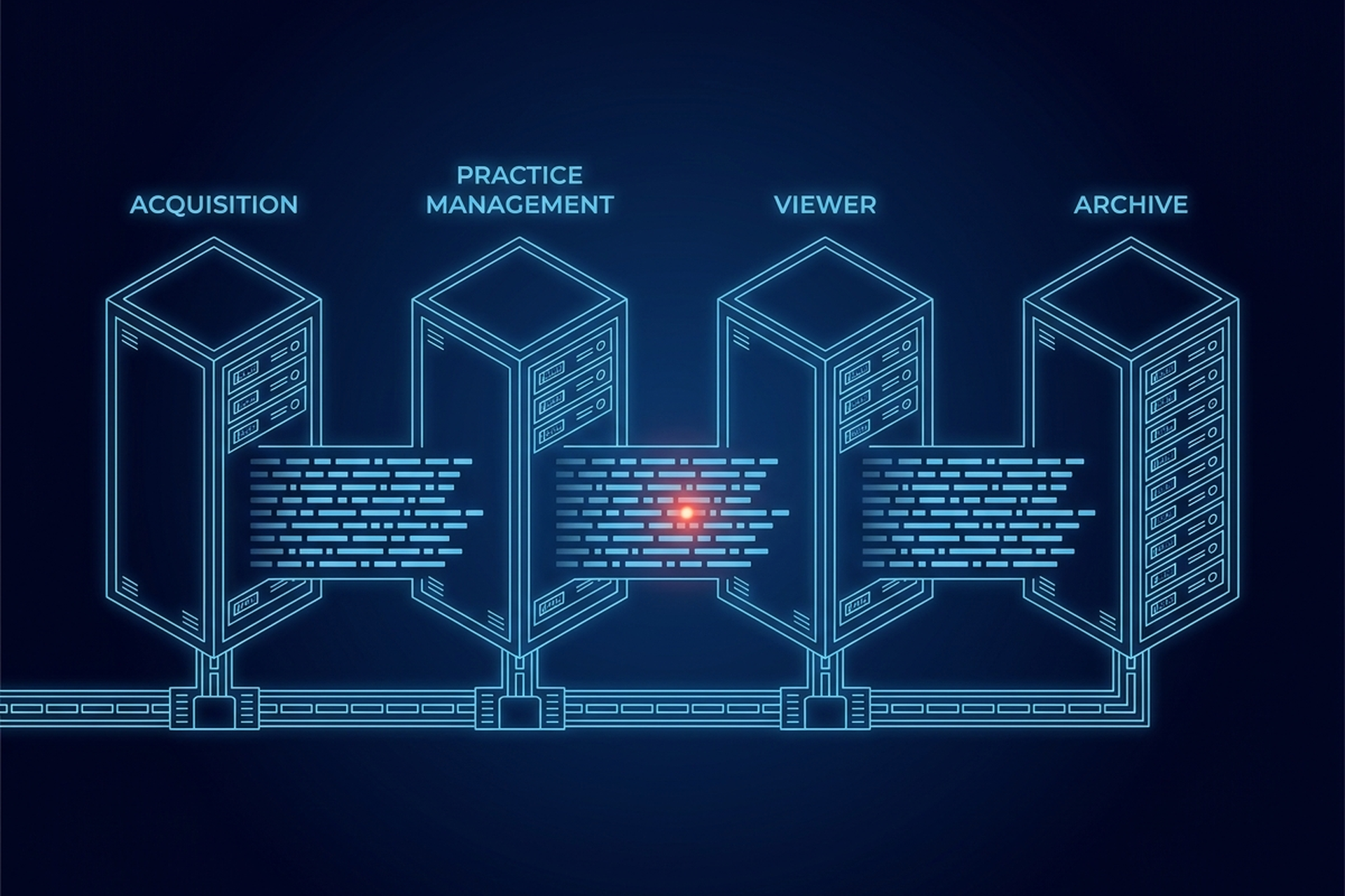
A bitewing exposure leaves the sensor, gets filed next to the patient’s chart, gets opened and measured by a clinician, and then sits in an archive for as long as the retention policy says. Four different pieces of software handle those four steps. All four are required, and none of them is the whole category on its own.
Acquisition software pulls the raw exposure off the sensor. A practice-management platform’s imaging module files it next to the chart. A dedicated viewer opens and measures it. A separate archive keeps the study retrievable long after the visit.
That runs against how the label usually gets sold. Sensor manufacturers built closed systems for years, bundling acquisition, viewing, and light record-keeping into one package tied to their own hardware. Buyers learned to treat dental imaging software as shorthand for whatever came in that box. The box has not disappeared, but the category underneath it has been formally divisible for longer than the marketing around it suggests.
The Four Layers Dental Imaging Software Groups Together
Four pieces make up the category, and a given product usually owns one or two of them rather than all four. Acquisition, or bridge, software talks directly to the sensor or plate reader and turns its output into a usable image file. A practice-management imaging module does that same job differently: it lives inside the scheduling, charting, and billing system, so the image lands next to the patient record automatically.
A dedicated viewer exists purely to open, measure, and annotate the image once it has been captured, independent of where it was stored. An archive keeps the finished study retrievable on its own timeline, separate from whichever system wrote it there first.
The Acquisition Layer
Acquisition software sits closest to the hardware. It receives whatever signal the sensor, plate reader, or panoramic unit produces and turns it into a displayable, storable file. How that resolution compares across an intraoral sensor, a panoramic sweep, or a cone beam volume is a format and display question, not a software-category one.
Many sensors ship with their own acquisition driver. Others rely on a shared, hardware-agnostic bridge that a practice-management system calls into, so one bridge can serve sensors from more than one manufacturer. That distinction, a proprietary driver versus a shared bridge, is the first thing an OEM or VAR has to identify before promising sensor compatibility, because the two paths carry different support and update obligations.
The Practice-Management Imaging Module
A practice-management system’s imaging module solves a different problem: keeping the image attached to the right patient, procedure code, and claim, without a separate login or a separate search. That convenience used to come at a cost. For years, a practice-management vendor’s imaging module worked only with the sensors that same vendor also sold, so choosing the records system also chose the hardware.
The same basic split shows up in general radiology, where a radiology information system owns the schedule and the record while a separate PACS owns the pixels. Dental practice-management platforms and their imaging modules divide the same way, under different names, and often with a far less consistent line between them.
That closed pairing has loosened industry-wide, though not uniformly. Many practice-management platforms now expose an open interface that a third-party acquisition tool can call into, so a practice can keep its records system and still change sensor brands. Where that interface does not exist, the imaging module and the hardware remain a single purchasing decision, whatever the label on the box says.
The Viewer Is Its Own Layer
A dental imaging viewer is built to open, measure, and annotate a study once it already exists. It does not need to know how the image was captured or where it will end up living long-term.
What a dental viewer has to get right, tooth numbering, magnification handling on a panoramic study, cone beam reformatting along the dental arch, is its own engineering problem. Whether a given product sold as dental imaging software includes one at all is a separate question. A product can excel at one and still leave the other three pieces of the category untouched.
The Archive Layer, and Where the FDA Draws Its Lines
The clearest evidence that dental imaging software divides into real, separable parts is regulatory, not commercial. The FDA does not treat medical imaging as one device. The software that manages and processes images has its own category, the hardware that stores them has another, and the dental x-ray system that made the exposure is regulated separately again. None of the three is defined by reference to the other two.
21 CFR 892.2050, classified Class II, covers a medical image management and processing system. Its identification paragraph describes “the review and digital processing of medical images for the purposes of interpretation by a trained practitioner of disease detection, diagnosis, or patient management.” That text names no modality and no practice-management system.
The line the regulator drew is between the software layer and the device that captured the study. It is the same line a complete platform holds for general radiology, just scoped to one specialty.
Underneath the classification sits the question a vendor neutral archive answers for a general-radiology buyer: whether the images outlive the system that first wrote them there, or disappear the day that vendor relationship ends.
What the ADA Told Regulators About the Seams
Splitting the category into acquisition, practice-management module, viewer, and archive is not just an engineering convenience. It is also where dental imaging goes wrong in practice. The American Dental Association told federal regulators in March 2026 that dental diagnostic images “are often stored in systems that operate independently of electronic dental records, restricting efficient exchange between dentists.” Its own explanation followed: “Proprietary software limits accessibility and interoperability necessary to coordinate patient-centered care and lack of standardization increases the cost of providing care.”
To inform that comment, the ADA convened a session with software vendors, imaging experts, clinicians, researchers, and standards specialists. The problems those participants raised, reliance on proprietary formats, inconsistent DICOM implementation, and fragmented exchange pathways, are symptoms of a familiar pattern. A study that leaves the acquisition layer cleanly can still be unreadable, or simply invisible, to whichever archive or practice-management system is supposed to receive it next.
Which of the Four a Dental Claim Covers
A claim of dental support is a claim about one or more of the four pieces, and the first question is which. The transport underneath all four does not change by modality. EBM mAIn PACS® lists native C-STORE, Query/Retrieve, Modality Worklist, and Storage Commitment as its published DICOM services. Those same services are what a dental acquisition tool, practice-management bridge, viewer, or archive would call on to move a study between systems.
Above that transport the four diverge, and each carries its own version of the question. On acquisition, the question is whether the layer handles every sensor brand already in use at the practice or only one manufacturer’s own hardware. On the practice-management side, it is whether an open interface exists or the imaging module is locked to a single hardware line. On the viewer, it is whether tooth numbering, panoramic magnification, and cone beam reformatting behave the way the practice already reads studies.
The archive question outlasts the other three. A study has to stay retrievable after the vendor relationship that first wrote it there ends. A product that clears the first three layers can still leave that one unanswered.
Assembling all four into one dental-specific product is a separate build from owning any single piece of it. A vendor that owns one layer can still be worth building around, provided the other three get sourced deliberately rather than assumed to come along with it.